Arch Iran Med. 28(6):367-369.
doi: 10.34172/aim.34254
Case Report
Bilateral Mydriasis in a Post-parotidectomy Patient: A Case Report
Ahmad Daneshi Conceptualization, Formal analysis, Investigation, Methodology, Supervision, Validation, Writing – review & editing, 1 
Saleh Mohebbi Conceptualization, Formal analysis, Investigation, Methodology, Supervision, Validation, Writing – review & editing, 1
Milad Shemshadi Conceptualization, Formal analysis, Investigation, Methodology, Validation, Writing – original draft, 1
Hadi Ghanbari Conceptualization, Formal analysis, Investigation, Methodology, Project administration, Supervision, Validation, Writing – review & editing, 1, *
Author information:
1Head & Neck Research Department, Hazrat Rasoul Hospital, Iran University of Medical Sciences, Tehran, Iran
Abstract
Anticholinergic agents, such as atropine and glycopyrrolate, are commonly utilized during anesthesia for their effects on secretion reduction and vagal activity. However, substantial dosages can induce mydriasis, which poses diagnostic challenges, particularly in head and neck surgeries. Despite their clinical relevance, limited studies explore these effects. A 35-year-old female presented with a left parotid mass and scheduled for a left superficial parotidectomy. Preoperatively, the patient exhibited normal ocular and neurological function. Postoperatively, fine bilateral ptosis, predominantly on the left side, and bilateral unresponsive mydriasis were noted. Anticholinergic-induced pupillary changes may mimic neurological pathology, underscoring the necessity for meticulous postoperative evaluation and awareness among clinicians.
Keywords: Case report, General anesthesia, Myasthenia gravis, Mydriasis
Copyright and License Information
© 2025 The Author(s).
This is an open-access article distributed under the terms of the Creative Commons Attribution License (
https://creativecommons.org/licenses/by/4.0), which permits unrestricted use, distribution, and reproduction in any medium, provided the original work is properly cited.
Cite this article as: Daneshi A, Mohebbi S, Shemshadi M, Ghanbari H. Bilateral mydriasis in a post-parotidectomy patient: a case report. Arch Iran Med. 2025;28(6):367-369. doi: 10.34172/aim.34254
Introduction
Sympathomimetic drugs containing local anesthetics or systemic anticholinergics are frequently used in different conditions and can cause unilateral or bilateral pupillary dilatation.1 Evidence suggests that the sympathetic nervous system does not mediate pupillary dilatation.2 anticholinergic drugs were widely used as a premedication in general anesthesia in order to reduce the secretion and vagal over activity and prevent bradycardia during the surgery. In addition, these drugs were used at the time of reversal of neuromuscular block.3 The administration of anticholinergic agents, particularly atropine and glycopyrrolate, at substantial dosages induces pupillary dilation (mydriasis) in adult patients who present with normal ocular function, with strabismus being the sole exception. This pharmacological response occurs through the blockade of parasympathetic nerve signals at muscarinic receptors in the iris sphincter muscle. It is noteworthy that when these medications are administered at standard therapeutic doses via intramuscular or intravenous routes, they typically do not elicit significant mydriatic effects. This dose-dependent relationship between anticholinergic drugs and pupillary response serves as an important clinical consideration in both anesthetic practice and emergency medicine.4 there is a diagnostic challenge due to the mydriatic effects of these drugs can interfere the neurological assessment after surgery. Despite their widespread use, there is limited studies exploring this side effect of anticholinergic-induced pupillary changes in surgeries, specifically in head and neck surgeries.
Case Report
The case was a 35-year-old female with no significant previous medical history that referred to our hospital clinic with the chief complaint of a left parotid mass that she noticed from 2 years ago, gradually increasing in size without any pain or skin involvement. In physical examination, she was a young female with lightly pigmented eyes and an asymmetrical face in the parotid area. A mass was palpated in the left parotid with a diameter of 2.5 cm × 3 cm with no sign of skin involvement or tenderness. The facial nerve examination was normal and symmetrical. She had done a CT-scan and MRI before, and parotid pleomorphic adenoma was confirmed by fine needle aspiration cytology (FNAC). According to this set of data, she was scheduled for left superficial parotidectomy. In the COVID-19 era, RT-PCR for COVID-19 was negative, and other laboratory tests were within normal limits (Table 1). In the operating room, lidocaine and epinephrine 1/100 000 were locally administered before skin incision. At the start of induction, total intravenous anesthesia was administered using thiopental 400 mg, atracurium 35 mg, fentanyl 15 mg, midazolam 2 mg, remifentanil 200 microgram, propofol 1% (300-400 mg total) during the operation time of approximately 70 min. The patient’s condition was reversed by atropine 1.5 mg and neostigmine 3 mg. The patient awakened slowly with no problem, but slower than the other patients with no facial nerve dysfunction in the recovery room. The patient received no antibiotic or other drugs locally or systemically in the operating room or after that. While visiting the patient post-operation in the ward, the senior surgeon noted a fine, barely detectable bilateral ptosis, predominantly on the left side. Recognizing bilateral unresponsive mydriasis in both eyes (Figure 1) and a quick overshooting upward movement followed by a downdrift of the upper lid was denoted as a positive Cogan’s sign. According to these findings, neurological disorders such as myasthenia gravis were suspected, and following imaging including Magnetic resonance imaging revealed no abnormal findings in the brain or brainstem, and pulmonary CT-scan was unremarkable. Acetylcholine receptor (AChR) antibody and COVID-19 serologic tests were requested. Immunological tests of COVID-19 (IgG and IgM) were negative, and the AChR antibody was positive, and the myasthenia gravis was confirmed (Table 2). In this case, we could consider the mydriasis as a side effect of the anti-cholinergic drugs used during general anesthesia, but precise examination and broader investigations helped us to detect the disease in its very early stage.
Table 1.
Baseline Preoperative Laboratory Values
|
Test
|
Result
|
Unit
|
Reference Value
|
| Hb |
12.8 |
g/dl |
Female: 12-16 |
| Plt |
226 |
*1000/mm3 |
140-440 |
| Blood Sugar |
96 |
mg/dL |
70-115 |
| B.U.N |
13 |
mg/dL |
5-23 |
| Creatinine |
0.8 |
mg/dL |
0.5-1.5 |
| INR |
1.08 |
Index |
1-1.1 |
| PTT |
31 |
Sec |
25-40 |
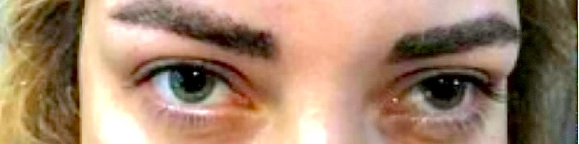
Figure 1.
Bilateral Mydriasis of Unknown Etiology After General Anesthesia for Left-Side Parotid Mass Resection
.
Bilateral Mydriasis of Unknown Etiology After General Anesthesia for Left-Side Parotid Mass Resection
Table 2.
Serological Test Result Supporting the Diagnosis of Myasthenia Gravis
|
Test
|
Result
|
Unit
|
Reference Value
|
| AChR antibody |
0.7 |
Nmol/L |
Negative < 0.4,
Borderline > 0.4- < 0.5,
Positive > 0.5 |
Discussion
Large doses of anticholinergic drugs (atropine, glycopyrrolate) produced mydriasis, though the usual intramuscular and intravenous doses of these drugs do not have this tendency. Neostigmine methyl-sulfate reduces the mydriatic effect when given intravenously in conjunction with atropine. Mydriasis was more likely to occur in lightly pigmented eyes.3 A 35-year-old female with a lightly pigmented eye was admitted to an outpatient clinic for parotidectomy. A meticulous history was taken from the patient, revealing the routine daily intake of propranolol 20 mg/d and levothyroxine 100 mg/d early in the morning of the operation day. No potential mydriatic effects were recognized before the injection of reverse drugs as a combination of atropine and Neostigmine. Volatile anesthesia was not used as isoflurane or sevoflurane, and bilateral mydriasis regression was delayed over 24 hours. Our case is not the same as that demonstrated utilizing inhalational drugs. could induce mydriasis1,5,6 There were no anticholinergic effects on increasing cardiac output or heart rate in our patient.7 Although patients with potentially increased intracranial pressure and mydriasis are often first investigated in the emergency room or intensive care unit, it is crucial to know the drugs that can be used as volatile anesthetics that may be able to induce mydriasis under exceptional circumstances. The patient was in good condition and had no symptoms of systemic atropine poisoning such as drowsiness, central nervous system depression, circulatory collapse with respiratory failure, sudden dizziness, headache, confusion, balance problems, or other symptoms of a possible stroke.8 Although Cogan’s lid twitch is not specific for ocular myasthenia gravis, but it should be considered.9 In our study, we faced a patient whose AChR antibody was more than the range, denotes myasthenia gravis with undetectable ptosis; Cogan’s positive sign and ocular presentation confirm the disease with the highest specificity 100%,10 which is rewarding for the patient and physician before presenting symptoms of the actual disease occur.
Conclusion
This case highlights the diagnostic complexity associated with bilateral mydriasis following anticholinergic use during general anesthesia, particularly in patients with lightly pigmented eyes. The persistence of mydriasis beyond the immediate postoperative period and the presence of a positive Cogan’s sign prompted a broader diagnostic evaluation, leading to the identification of ocular myasthenia gravis with high specificity. This underscores the importance of recognizing subtle ocular signs as potential indicators of underlying neuromuscular conditions, which could be critical for early diagnosis and management. Clinicians should maintain a high index of suspicion for non-neurological causes of mydriasis while also considering systemic implications, especially during routine anesthetic practice.
Competing Interests
The authors declare no competing interests.
Ethical Approval
This research was conducted with the approval of the Ethics Committee of Iran University of Medical Sciences, following all relevant ethical guidelines and regulations. Written informed consent was obtained from the patient.
Funding
This work is not supported by any funding from a company or granting agency, nor is it paid to the authors. Equipment explicitly used for the case described is also not billed to the patient, and laboratory tests are not billed to the patient. Support was provided solely from institutional and departmental sources.
References
- von Arx T, Lozanoff S, Zinkernagel M. [Ophthalmologic complications and local anesthesia Pathophysiology and types of eye complications after intraoral dental anesthesia, and clinical recommendations]. Swiss Dent J 2014; 124(11):1189-96. doi: 10.61872/sdj-2014-11-02.[German] [Crossref] [ Google Scholar]
- Nowak S, Rehberg S, Schroeder HWS, Fleck S. Pupillary dilatation associated with volatile anaesthetics: a small case series. Br J Anaesth 2017; 119(1):169-70. doi: 10.1093/bja/aex183 [Crossref] [ Google Scholar]
- Mirakhur RK, Clarke RS, Dundee JW, McDonald JR. Anticholinergic drugs in anaesthesia A survey of their present position. Anaesthesia 1978; 33(2):133-8. doi: 10.1111/j.1365-2044.1978.tb08340.x [Crossref] [ Google Scholar]
- Schwartz H, Apt L. Mydriatic effect of anticholinergic drugs used during reversal of nondepolarizing muscle relaxants. Am J Ophthalmol 1979; 88(3 Pt 2):609-12. doi: 10.1016/0002-9394(79)90523-3 [Crossref] [ Google Scholar]
- Bomberg H, Groesdonk HV, Bellgardt M, Volk T, Meiser A. AnaConDaTM and MirusTM for intensive care sedation, 24 h desflurane versus isoflurane in one patient. Springerplus 2016; 5:420. doi: 10.1186/s40064-016-2065-0 [Crossref] [ Google Scholar]
- Purrucker JC, Renzland J, Uhlmann L, Bruckner T, Hacke W, Steiner T. Volatile sedation with sevoflurane in intensive care patients with acute stroke or subarachnoid haemorrhage using AnaConDa®: an observational study. Br J Anaesth 2015; 114(6):934-43. doi: 10.1093/bja/aev070 [Crossref] [ Google Scholar]
- Larson MD, Tayefeh F, Sessler DI, Daniel M, Noorani M. Sympathetic nervous system does not mediate reflex pupillary dilation during desflurane anesthesia. Anesthesiology 1996; 85(4):748-54. doi: 10.1097/00000542-199610000-00009 [Crossref] [ Google Scholar]
- North RV, Kelly ME. A review of the uses and adverse effects of topical administration of atropine. Ophthalmic Physiol Opt 1987; 7(2):109-14. doi: 10.1111/j.1475-1313.1987.tb01004.x [Crossref] [ Google Scholar]
- Nair AG, Patil-Chhablani P, Venkatramani DV, Gandhi RA. Ocular myasthenia gravis: a review. Indian J Ophthalmol 2014; 62(10):985-91. doi: 10.4103/0301-4738.145987 [Crossref] [ Google Scholar]
- Padua L, Stalberg E, LoMonaco M, Evoli A, Batocchi A, Tonali P. SFEMG in ocular myasthenia gravis diagnosis. Clin Neurophysiol 2000; 111(7):1203-7. doi: 10.1016/s1388-2457(00)00307-2 [Crossref] [ Google Scholar]